Video Credit: Ben Brandon, 2022 Summer Intern
Alzheimer's disease is one cause of dementia. Historically, it is a disease of the brain which results in impairment of cognitive brain functions, particularly memory and learning.
For over a hundred years, we've known that the characteristic anatomical findings in the brain of someone with Alzheimer's disease include generalized atrophy, or the wasting away, of the brain. In Alzheimer's disease, the shrinkage can be seen in the parts of the brain that are central to memory and learning.
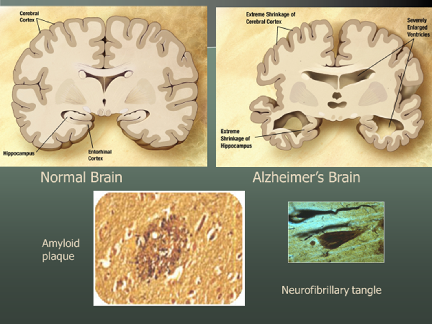 When viewed under a microscope, there are two major types of lesions in a brain with Alzheimer's. They are glue-like amyloid plaques and abnormal tangles of a protein called tau that collects inside neurons. Beta-amyloid plaques appear as dark clumps collecting between neurons. The tangles (tau) look like very dark, or black, long pear shapes.
These findings were first described by Alois Alzheimer at the beginning of the 20th century. Since then, we've learned a great deal about cell biology and biochemistry which leads to the development of plaques and tangles. It is summarized in this cartoon below. Within the bubble is what happens in Alzheimer's disease and outside the bubble is the normal process.

The row of pink balls depicts the cell membrane of a neuron. There are many structures within the cell membrane. There are also many organelles and many cells within the brain structure. Shown outside the bubble are the normally functioning structures such as the microtubules (that help transport materials around the neuron) and the mitochondria (which produce energy so that the cell can stay alive and function). The cell membrane itself contains proteins and channels which enable the internal cell to interact safely and effectively with its environment while maintaining cell integrity.
In Alzheimer's disease, depicted inside the bubble, things change. A normal protein crossing the cell membrane is abnormally clipped. That releases a fragment that is shown as that orange strand between the two clippers within the bubble. That is a fragment of a normal protein, but that protein is highly toxic. It's very sticky. It forms little aggregates of Beta-amyloid. This is now shown in the upper left part of the bubble. These aggregates are called oligomers and the oligomers condense to form fibrils. Then they deposit as plaques. Once the plaques deposit, other things happen, and neurons become damaged. Ultimately all of the organelles within the neuron, including the microtubules and the mitochondria start to disintegrate, dysfunction, and tangles start to form within themselves.